RSVP Phantom™
Radiosurgery Verification Phantom
The RSVP Phantom™ was developed to provide stereotactic localization and dose verification for radiosurgery machines. The phantom may be used for a variety of radiosurgery applications, including periodic quality assurance evaluations and acceptance testing. In addition, the phantom may be used to perform
re-evaluations after equipment and software upgrades.
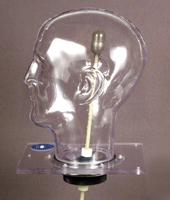
The phantom's design provides full simulation of the localization and irradiation sequences. The anatomically accurate head form is filled with water to simulate the radiation absorption and scatter of human soft tissue. The heavy-duty outer shell is designed to accommodate the anchoring screws.
An internal container called a tumor vessel can be positioned anywhere within the head form by manipulating an external position rod. This vessel may be filled with a radiation-sensitive gel for alignment evaluations or with TL dosimeters for quantitative dose measurements. An Exradin Microchamber may also be used with a special optional chamber holder and ball assembly.
Stereotactic Localization - An agarose gel that changes color after exposure to a minimum radiation dose of 40 Gy is used. A small radio-opaque target is placed inside the gel. Next, the tumor vessel is positioned within the head form and the radiosurgery head frame is attached to the phantom. The standard protocols are followed to locate the x, y and z coordinates of the patient's tumor through angiography, CT, or MR imaging. The phantom is then mounted in the radiosurgery system and irradiated. The gel is carefully extracted, sliced and analyzed to determine whether the location and shape of the irradiated portion of the gel corresponds to the desired target location.
Dose Measurements - Either TLD or radiochromic film may be used with appropriate holders to quantify the radiation dose. The phantom can also accommodate a specially designed chamber holder for the Exradin Microchamber Model A14.
Construction - The shell of the RSVP Phantom™ is formed from a transparent, 1/4" cellulose acetate butyrate sheet, chosen for its strength and low water absorption. The shell is mounted on a polycarbonate end plate, and the tumor port and cover plate assembly are attached with nylon screws to the end plate. The cover plate is removable for internal access. The tumor vessel is attached to the external position rod. After the desired position is reached, the vessel is locked into place by hand-tightening a lock nut on the rotation ball and a lock bolt on the position rod. The RSVP Phantom™ includes three tumor vessels and a wooden storage case.